Dental X-Ray Positioning Guide
This guide offers a comprehensive overview of dental X-ray positioning techniques. It will assist dental professionals in capturing high-quality radiographic images. Proper positioning ensures accurate diagnoses, and effective treatment planning for various dental conditions.
Dental radiography is a crucial diagnostic tool in modern dentistry, providing valuable insights into oral health beyond what can be seen during a clinical examination. X-rays, or radiographs, enable dentists to detect hidden dental structures, cavities, bone loss, and malignant masses. They serve as an adjunct to clinical exams for proper diagnosis of dental diseases.
Intraoral and extraoral techniques are used to capture detailed images of teeth and surrounding tissues. Understanding the principles and techniques of dental radiography is essential for every dental professional. Accurate positioning of the patient, film or sensor, and X-ray beam are vital for obtaining diagnostic radiographs. Proper processing techniques are also important for creating accurate images.

This guide will explore the basics of dental radiography, including equipment, types of radiographs, and essential positioning techniques; It will also address common errors and corrections to help dental professionals enhance their skills and ensure patient safety while capturing the best images.
Types of Dental X-Rays
Dental X-rays are categorized into two main types⁚ intraoral and extraoral. Intraoral X-rays, taken inside the mouth, provide detailed views of individual teeth and surrounding structures. Extraoral X-rays, taken outside the mouth, offer broader views of the jaws and skull.
Intraoral radiographs include bitewing and periapical X-rays. Bitewing X-rays focus on the crowns of the upper and lower teeth, helping to detect cavities and assess bone levels. Periapical X-rays show the entire tooth, from crown to root, along with surrounding bone, aiding in the diagnosis of root problems and infections.
Extraoral radiographs include panoramic and cephalometric X-rays, as well as Cone Beam Computed Tomography (CBCT). Panoramic X-rays provide a single image of the entire mouth, while cephalometric X-rays capture an image of the entire head. CBCT offers a 3D view of the teeth and surrounding structures. Each type serves a unique purpose in comprehensive dental diagnostics.
Intraoral X-Rays
Intraoral X-rays are a crucial diagnostic tool in dentistry, providing detailed images of teeth, bone, and surrounding structures. These X-rays are taken with the film or digital sensor placed inside the patient’s mouth, allowing for high-resolution views of specific areas of concern; The primary types of intraoral X-rays include bitewing and periapical radiographs.
Bitewing X-rays are primarily used to detect interproximal caries, or cavities between the teeth, and to assess the height of the alveolar bone, indicating potential bone loss due to periodontal disease. Periapical X-rays, on the other hand, offer a complete view of the tooth, from the crown to the root, and the surrounding bone.
These are essential for diagnosing apical lesions, root fractures, and other abnormalities affecting the tooth’s apex. Proper technique in taking intraoral X-rays, including correct film placement and beam angulation, is crucial for obtaining accurate and diagnostic images.
Bitewing Radiography
Bitewing radiography is a common intraoral technique used to visualize the crowns of the upper and lower posterior teeth simultaneously. The primary purpose of bitewing X-rays is to detect interproximal caries, assess the marginal ridge relationship, and evaluate the alveolar bone levels. During the procedure, a film or digital sensor is positioned parallel to the crowns of the teeth.
The patient then bites down on a tab or bitewing holder to stabilize the receptor. The central ray of the X-ray beam is directed through the contacts of the teeth with a slight vertical angulation. Correct horizontal angulation prevents overlapping of the teeth, ensuring clear visualization of the contact areas.
Bitewing radiographs are an essential tool for early detection of dental decay and monitoring the progression of periodontal disease. They are a routine part of dental examinations, aiding in proactive dental care.
Periapical Radiography
Periapical radiography is an intraoral X-ray technique that provides a complete view of a tooth, from the crown to the apex, and the surrounding bone. This type of radiograph is essential for assessing the health of the root, detecting periapical lesions, and evaluating bone support. Two main techniques are employed for periapical radiographs⁚ the paralleling technique and the bisecting angle technique.
The paralleling technique involves positioning the film receptor parallel to the long axis of the tooth, using a film holder to maintain this position. The central X-ray beam is directed perpendicular to both the tooth and the receptor. This method minimizes distortion and provides a more accurate representation of the tooth’s length and surrounding structures.
Periapical radiographs are crucial for diagnosing apical infections, evaluating root fractures, and assessing the outcome of endodontic treatments. They are invaluable for comprehensive dental evaluations.
Extraoral X-Rays
Extraoral X-rays are radiographic images taken with the film or sensor positioned outside the patient’s mouth. These radiographs offer a broader view of the oral and maxillofacial structures, making them valuable for assessing overall dental health and identifying abnormalities beyond individual teeth.
Unlike intraoral X-rays that focus on specific teeth or small areas, extraoral X-rays provide a comprehensive overview of the jaws, temporomandibular joints (TMJ), sinuses, and other facial structures. Common types of extraoral X-rays include panoramic radiographs, cephalometric radiographs, and cone beam computed tomography (CBCT). Panoramic radiographs provide a wide view of the entire dentition and surrounding structures on a single image. Cephalometric radiographs are used to evaluate the skeletal relationships of the jaws and skull, often used in orthodontics. CBCT provides three-dimensional images.
Extraoral X-rays are essential diagnostic tools in various dental specialties.

Panoramic Radiography
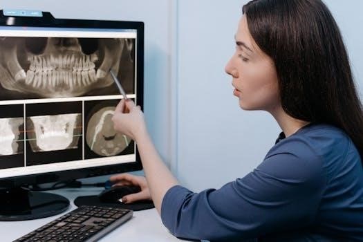
Panoramic radiography, also known as a panoramic X-ray, is an extraoral imaging technique that provides a broad view of the entire dentition, including the upper and lower jaws, temporomandibular joints (TMJs), and surrounding structures, all on a single image. This type of radiograph is commonly used in dental practice for a variety of diagnostic purposes.
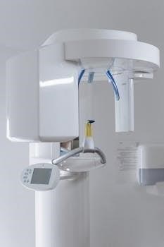
During a panoramic X-ray, the patient stands or sits still while the X-ray machine rotates around their head. The X-ray beam passes through the patient’s head, and the image is captured on a film or digital sensor. The resulting image displays a flattened, two-dimensional representation of the curved dental arches and related anatomical structures.
Panoramic radiographs are valuable for assessing impacted teeth, jaw fractures, tumors, cysts, and other abnormalities that may not be visible on intraoral X-rays. They are also useful for evaluating the development of teeth in children and adolescents. However, panoramic radiographs may not provide the same level of detail as intraoral X-rays for detecting small cavities.
Cephalometric Radiography
Cephalometric radiography is an extraoral imaging technique primarily used in orthodontics to evaluate the skeletal and soft tissue relationships of the head and face. It provides a standardized lateral view of the skull, allowing for precise measurements and analysis of craniofacial structures.
During cephalometric radiography, the patient’s head is positioned in a cephalostat, a device that stabilizes the head and ensures consistent positioning for reproducible images. The X-ray beam passes through the patient’s head from the side, and the image is captured on a film or digital sensor.
Cephalometric radiographs are essential for orthodontic treatment planning, monitoring growth and development, and assessing the effectiveness of orthodontic treatment. They are also used in orthognathic surgery planning to evaluate skeletal discrepancies and predict surgical outcomes. Cephalometric analysis involves measuring angles and distances between specific anatomical landmarks on the radiograph to assess skeletal and dental relationships.

These measurements help orthodontists diagnose malocclusions, determine the appropriate treatment approach, and track changes during treatment.
Cone Beam Computed Tomography (CBCT)
Cone Beam Computed Tomography (CBCT) is an advanced extraoral imaging technique that provides three-dimensional images of the maxillofacial region. Unlike traditional two-dimensional radiographs, CBCT offers detailed visualization of bone, teeth, and soft tissues, allowing for more accurate diagnoses and treatment planning.
During a CBCT scan, the patient sits or stands while an X-ray beam rotates around their head, capturing multiple images from different angles. These images are then reconstructed using computer software to create a three-dimensional volume of the scanned area.
CBCT is valuable for various dental applications, including implant planning, endodontic assessment, oral and maxillofacial surgery, and the evaluation of temporomandibular joint disorders. It allows clinicians to assess bone density, identify anatomical variations, and detect pathological conditions with greater precision than traditional radiographs.
The use of CBCT should be justified based on the patient’s specific clinical needs, considering the radiation dose and potential benefits. Proper training and adherence to ALARA principles are essential to minimize radiation exposure while maximizing diagnostic information.

Intraoral Radiography Techniques
Intraoral radiography techniques involve placing the X-ray film or digital sensor inside the patient’s mouth to capture detailed images of the teeth and surrounding structures. These techniques are essential for diagnosing various dental conditions, including caries, periodontal disease, and periapical lesions.
Two primary intraoral radiography techniques are commonly used⁚ the paralleling technique and the bisecting angle technique. The paralleling technique aims to position the film or sensor parallel to the long axis of the tooth, ensuring minimal distortion and accurate representation of the tooth’s dimensions. This technique typically requires the use of a film holder or positioning device to maintain parallelism.
The bisecting angle technique, on the other hand, involves positioning the film or sensor as close as possible to the tooth while bisecting the angle formed by the tooth’s long axis and the film plane. The X-ray beam is then directed perpendicular to the bisector. Although simpler to execute, this technique is more prone to distortion and may not provide as accurate an image as the paralleling technique.
Paralleling Technique
The paralleling technique is a cornerstone of intraoral radiography, aiming to minimize distortion and maximize accuracy in dental X-ray images. This technique involves positioning the X-ray film or digital sensor parallel to the long axis of the tooth being radiographed. This parallelism is crucial for achieving a true representation of the tooth’s length and shape.
To achieve parallelism, specialized film holders or positioning devices are used. These devices help maintain the film or sensor’s position, ensuring it remains parallel to the tooth even when anatomical constraints, such as a shallow palate, are present. The X-ray beam is then directed perpendicularly to both the tooth and the film or sensor.
The paralleling technique offers several advantages, including reduced distortion, improved image clarity, and more accurate measurements. However, it may require careful patient positioning and the use of specialized equipment, making it slightly more complex than other techniques.
Bisecting Angle Technique
The bisecting angle technique is an alternative intraoral radiographic method used when paralleling is difficult. This technique relies on an imaginary bisector, which is a line that divides the angle formed by the tooth’s long axis and the film or sensor plane into two equal angles. The X-ray beam is then directed perpendicularly to this imaginary bisector.
Unlike the paralleling technique, the bisecting angle technique does not require the film or sensor to be parallel to the tooth. This makes it useful in situations where anatomical limitations, such as a shallow palate or tori, prevent proper placement of the film holder. However, the bisecting angle technique is more prone to distortion.
Careful attention to angulation is critical to minimize image distortion. Errors in vertical angulation can result in foreshortening or elongation of the tooth image, while errors in horizontal angulation can cause overlapping of adjacent structures. Despite its limitations, the bisecting angle technique remains a valuable tool.
Patient and Equipment Positioning
Proper patient and equipment positioning are crucial for obtaining diagnostic-quality dental radiographs while minimizing radiation exposure. The patient should be seated comfortably in the dental chair with their head stabilized using a headrest. The midsagittal plane should be perpendicular to the floor, and the occlusal plane should be parallel to the floor unless specific angulation is required.
The X-ray tube head must be positioned accurately according to the chosen radiographic technique (paralleling or bisecting angle). For intraoral radiographs, the position indicating device (PID) should be aligned to direct the central ray towards the center of the film or sensor. The PID should be as long as possible to reduce radiation exposure to the patient.
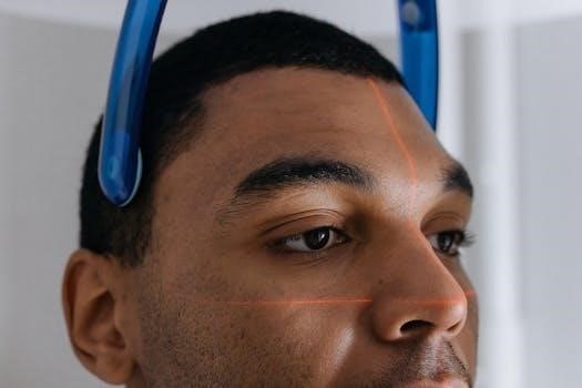
For extraoral radiographs like panoramic or cephalometric X-rays, the patient must be positioned according to the manufacturer’s instructions. This usually involves using head stabilizers and bite guides to ensure proper alignment and prevent movement during the scan; Precise positioning prevents artifacts.
Common Errors and Corrections
Several common errors can occur during dental radiography, affecting image quality and diagnostic accuracy. One frequent mistake is cone-cutting, where the X-ray beam doesn’t fully cover the film or sensor, resulting in a partially exposed image. This can be avoided by ensuring the PID is properly aligned and centered.
Another error is elongation or foreshortening of teeth, which occurs when the vertical angulation is incorrect during the bisecting angle technique. To correct this, adjust the vertical angulation according to the bisecting angle principle. Blurred images can result from patient movement during exposure. Instruct patients to remain still and use head stabilization devices when necessary.
Incorrect horizontal angulation can cause overlapping of teeth, hindering the detection of interproximal caries. Adjust the horizontal angulation to ensure the X-ray beam passes through the interproximal spaces. Overexposed or underexposed images can be corrected by adjusting the exposure settings on the X-ray machine.
